Health Assessment - Cardiovascular
Cardiovascular
The cardiovascular function domain in TILDA comprises objective physiological assessments designed to evaluate cardiovascular health, vascular function, and haemodynamic regulation in older adults. Measures collected across waves include blood pressure, pulse wave velocity, pulse wave analysis, beat-to-beat haemodynamic monitoring, electrocardiography (ECG), and heart rate variability, providing detailed information on cardiac function, arterial stiffness, autonomic regulation, and cardiovascular ageing.
Cardiovascular Measures
Click each heading to expand details of this test.
Overview
Measuring: Blood Pressure is measured as part of the Health Assessment.
Equipment used: Omron M10-IT
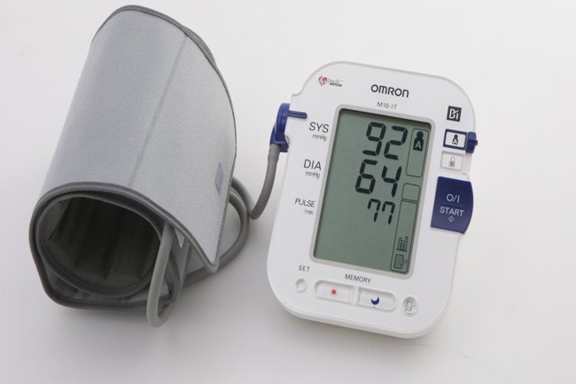
Equipment Description: The Omron M10-IT is a digital automated oscillometric BP monitor. BP measurements are taken from the upper arm using an arm cuff (22-42cm).
Assessment Type: Centre and Home Assessment
Protocol
- Measurements were taken according to a standard protocol at an ambient temperature of 20–25°C.
- BP was measured in the upper arm, at heart height, while the respondent was seated comfortably in an upright position.
- BP was recorded twice while seated with a timed interval of 1 min between readings.
- The mean systolic and diastolic readings were obtained from these two measurements.
- A third measurement is then taken immediately on standing form sitting, to capture postural drop.
- Standing heart rate, systolic and diastolic blood pressure measurements are obtained from this measurement.
Collection Waves
| Measure | Wave 1 | Wave 2 | Wave 3 | Wave 4 | Wave 5 | Wave 6 |
|---|---|---|---|---|---|---|
| Blood Pressure | X | X | X |
Example TILDA Papers
- McGarvey, C., Scarlett, S., Briggs, R., McCrory, C., McEvoy, J. W., Kenny, R. A., & Sexton, D. J. (2025). Adherence to the European Society of Cardiology hypertension guidelines over 12 years of follow-up in the Irish population. Open Heart, 12(2).
- Doyle, K., Rice, C. P., Kenny, R. A., Briggs, R., & Lavan, A. H. (2026). The effect of fludrocortisone and midodrine on ambulatory blood pressure biomarkers and symptoms of syncope. Blood Pressure, 35(1). https://doi.org/10.1080/08037051.2026.2618319
- O Regan, C., Kearney, P. M., Cronin, H., Savva, G. M., Lawlor, B. A., & Kenny, R. (2013). Oscillometric measure of blood pressure detects association between orthostatic hypotension and depression in population based study of older adults. BMC psychiatry, 13(1), 266.
Overview
Measuring: Heart rate variability (HRV) assesses autonomic nervous system function. Low HRV is considered an independent marker of mortality risk. Beat-to-beat variation is captured via ECG and must reflect true physiological variation, not measurement error.
Equipment used: - Medilog Darwin AR12 using Medilog Darwin software (waves 1 and 3) - Bittium using cardiscope software (Wave 6)
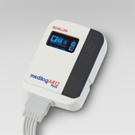
Medilog Darwin AR12 device
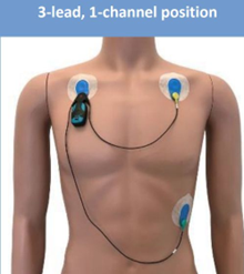
Bittium device
Equipment Description: ECG electrodes are attached to the participant and connected to the recording device. The software calculates R–R intervals from the ECG signal, from which HRV parameters are derived in both time and frequency domains. Raw ECG is visually inspected by trained staff to identify arrhythmias such as atrial fibrillation. Room temperature is maintained at 21–23 °C, and the participant is kept warm, particularly the finger attached to the Finometer cuff.
Assessment Type: Centre Assessment Only
Protocol
- Electrodes are applied and connected to the device, and the participant is positioned supine and allowed to rest for 5 minutes while breathing naturally.
- HRV is recorded for 5 minutes at the participant’s normal breathing rate, followed by 5 minutes of paced breathing guided by pre-recorded instructions.
- The participant is monitored throughout, and the test is stopped if discomfort occurs or the signal quality is compromised.
Collection Waves
| Assessment | Wave 1 | Wave 2 | Wave 3 | Wave 4 | Wave 5 | Wave 6 |
|---|---|---|---|---|---|---|
| Heart Rate Variability | X | X | X |
Example TILDA Papers
O’Regan C, Kenny RA, Cronin H, Finucane C, Kearney PM. Antidepressants strongly influence the relationship between depression and heart rate variability: findings from The Irish Longitudinal Study on Ageing (TILDA). Psychol Med. 2015 Feb;45(3):623-36. doi: 10.1017/S0033291714001767. Epub 2014 Jul 30. PMID: 25075912; PMCID: PMC4413849.
Frewen J, Finucane C, Savva GM, Boyle G, Coen RF, Kenny RA. Cognitive function is associated with impaired heart rate variability in ageing adults: the Irish longitudinal study on ageing wave one results. Clin Auton Res. 2013 Dec;23(6):313-23. doi: 10.1007/s10286-013-0214-x. PMID: 24077752.
McNicholas T, Tobin K, O’Callaghan S, Kenny RA. Is orthostatic hypotension more common in individuals with atrial fibrillation?-Findings from The Irish Longitudinal Study on Ageing (TILDA). Age Ageing. 2017 Nov 1;46(6):1006-1010. doi: 10.1093/ageing/afx096. PMID: 28985288.
Armstrong R, Wheen P, Brandon L, Finucane C, Kenny RA, Maree A. Impaired autonomic function after incomplete revascularisation. Open Heart. 2021 Oct;8(2):e001835. doi: 10.1136/openhrt-2021-001835. PMID: 34635578; PMCID: PMC8506863.
Overview
Measuring: : NIRS measures changes in oxygenated ([O₂Hb]) and deoxygenated ([HHb]) haemoglobin in the frontal lobe. This provides a non-invasive measure of cerebral perfusion during lying and standing, helping assess the effect of blood pressure changes on brain oxygenation.
Equipment used: PortaLite device using Oxysoft software.
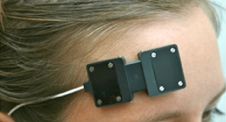
Equipment Description: The device records [O₂Hb], [HHb], and TSI continuously. A small optical sensor is placed on the participant’s forehead, secured with adhesive, which ensures stability. A black headband is placed over the sensor to prevent interference from ambient light.
Assessment Type: Centre Assessment Only
Protocol
- The participant is briefed and checked for adhesive allergies.
- The sensor is placed on the forehead and secured with adhesive and a headband, with the battery pack positioned safely.
- The PortaLite device is started via Oxysoft, and continuous measurements of oxy- and deoxyhaemoglobin, and Tissue Saturation Index (TSI), are recorded during rest, paced breathing, and the active stand.
- Key periods are marked in the software, and the participant is monitored throughout.
- The test is stopped if there is discomfort or signal loss, after which the device is removed and cleaned.
Collection Waves
| Measure | Wave 1 | Wave 2 | Wave 3 | Wave 4 | Wave 5 | Wave 6 |
|---|---|---|---|---|---|---|
| Near Infrared Spectoscropy | X | X |
Example TILDA Papers:
- Leidhin CN, McMorrow J, Carey D, Newman L, Williamson W, Fagan AJ, Chappell MA, Kenny RA, Meaney JF, Knight SP. Age-related normative changes in cerebral perfusion: Data from The Irish Longitudinal Study on Ageing (TILDA). Neuroimage. 2021 Apr 1;229:117741. doi: 10.1016/j.neuroimage.2021.117741. Epub 2021 Jan 14. PMID: 33454406.
- Newman L, Nolan H, Carey D, Reilly RB, Kenny RA. Age and sex differences in frontal lobe cerebral oxygenation in older adults-Normative values using novel, scalable technology: Findings from the Irish Longitudinal Study on Ageing (TILDA). Arch Gerontol Geriatr. 2020 Mar-Apr;87:103988. doi: 10.1016/j.archger.2019.103988. Epub 2019 Nov 18. PMID: 31775091.
- Maasakkers CM, Thijssen DH, Knight SP, Newman L, O’Connor JD, Scarlett S, Carey D, Buckley A, McMorrow JP, Leidhin CN, Feeney J, Melis RJ, Kenny RA, Claassen JA, Looze C. Hemodynamic and structural brain measures in high and low sedentary older adults. J Cereb Blood Flow Metab. 2021 Oct;41(10):2607-2616. doi: 10.1177/0271678X211009382. Epub 2021 Apr 17. PMID: 33866848; PMCID: PMC8504407.
Overview
Measuring: Phasic blood pressure allows for accurate assessment of dynamic blood pressure and is used to measure blood pressure responses during lying and standing. Continuous blood pressure is measured at the finger and generates a pressure waveform. The software derives brachial pressure waveforms and calculates multiple beat-to-beat haemodynamic parameters, all obtained during the active stand.
Equipment used: Finometer using BeatScope software.
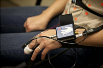
Equipment Description: Finometer: Beat-to-beat parameters: - Systolic and Diastolic BP and Mean Arterial Pressure (MAP) (mmHg) - Heart Rate (HR) (bpm) - Stroke Volume (SV) (mL) - Cardiac Output (CO) (L/min) - Total Peripheral Resistance (TPR) (dyn.s/cm5). - Left Ventricular Ejection Time (LVET) (ms) - Pulse Interval (PI) (ms)
Standing time (s), defined as the time taken by the participant to stand during the active stand, is also recorded.
Assessment Type: Centre Assessment Only
Protocol
- The participant is fitted with a finger cuff on the middle finger connected to the Finometer device and allowed to rest in a supine position.
- The arm is supported in a sling, with the hand resting across the chest just below the ribcage.
- Continuous beat-to-beat blood pressure is recorded during lying and throughout an active stand.
- The participant is instructed to stand promptly when directed, and phasic blood pressure and derived haemodynamic parameters are recorded using BeatScope software.
- Reassurance is provided throughout, and the test is discontinued if the participant experiences discomfort.
Collection Waves
| Measure | Wave 1 | Wave 2 | Wave 3 | Wave 4 | Wave 5 | Wave 6 |
|---|---|---|---|---|---|---|
| Phasic Blood Pressure | X | X | X |
Example TILDA Papers
- Hernández B, Dyer AH, Finucane C, Nipoti B, Romero-Ortuno R, Reilly R, Kenny RA. The Impact of Type 2 Diabetes on Peripheral and Cerebral Hemodynamic Responses to Active Stand. J Gerontol A Biol Sci Med Sci. 2024 May 1;79(5):glae073. doi: 10.1093/gerona/glae073. PMID: 38436476; PMCID: PMC11025558.
- A Shirsath M, O’Connor JD, Boyle R, Newman L, Knight SP, Hernandez B, Whelan R, Meaney JF, Kenny RA. Slower speed of blood pressure recovery after standing is associated with accelerated brain aging: Evidence from The Irish Longitudinal Study on Ageing (TILDA). Cereb Circ Cogn Behav. 2024 Feb 24;6:100212. doi: 10.1016/j.cccb.2024.100212. PMID: 38445293; PMCID: PMC10912350.
- McCrory C, Berkman LF, Moore PV, Kenny RA. What Explains Socioeconomic Differences in the Speed of Heart Rate Recovery to Postural Challenge? J Gerontol A Biol Sci Med Sci. 2017 Nov 9;72(12):1717-1723. doi: 10.1093/gerona/glx060. Erratum in: J Gerontol A Biol Sci Med Sci. 2017 Sep 1;72(9):1303. doi: 10.1093/gerona/glx095. PMID: 28407037.
Overview
Measuring: Pulse wave velocity is an indicator of arterial stiffness and is used as a surrogate marker for atherosclerosis. This measures the velocity of the blood travelling through the arteries (higher speed -> higher stiffness).
Equipment used: Vicorder device.
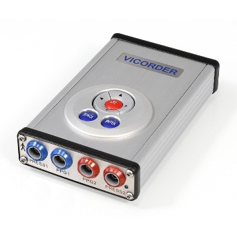
Equipment Description: This device measures carotid-femoral pulse wave velocity (value in m/s). It is necessary to measure the distance between the sternal notch and the femoral cuff beforehand. A total of two measurements are taken and then an average is calculated.
Assessment Type: Centre Assessment Only
Protocol
- The participant is positioned supine for measurement of the distance between the supra-sternal notch and the midpoint of the left thigh cuff.
- The thigh cuff is applied high on the upper left thigh and the neck cuff is positioned over the right carotid artery.
- The participant is then positioned at approximately 45° and two carotid–femoral pulse wave velocity measurements are taken using the Vicorder device.
- Measurements are recorded and the average pulse wave velocity (m/s) is calculated.
- Reassurance is provided throughout and the test is stopped if the participant experiences discomfort.
Collection Waves
| Measure | Wave 1 | Wave 2 | Wave 3 | Wave 4 | Wave 5 | Wave 6 |
|---|---|---|---|---|---|---|
| Pulse Wave Velocity | X | X | X |
Example TILDA Papers
- Horan M, Carey D, Knight S, Fagan A, Meaney JFM, Kenny RA, De Looze C. Examining the independent and moderating effects of arterial stiffness and cerebral blood flow on total hippocampal and hippocampal subfield volumes. Front Aging Neurosci. 2025 Jun 30;17:1466294. doi: 10.3389/fnagi.2025.1466294. PMID: 40671789; PMCID: PMC12264913.